Category Archives: Cases

Sleuth-It Diagnosis: Bicuspid aortic valve

[button label=”Read the Sleuth-It” url=”/sleuth-it-what-counts-in-the-heart” target=”_blank” color=”#f05134″]

Safe bicuspid

What counts in the heart? The number of parts to each valve counts. In this case, the number of parts (leaflets) of the aortic valve was important.

You stopped counting after seeing two aortic valve leaflets. Normally there are three.

Two aortic valves rather than three is an inherited condition called “bicuspid aortic valve.” It affects 1-2% of the population.

Bicuspid aortic valve can be associated with aortic aneurysm and rupture, typically in late middle age among males with the condition.

The inheritance is not well-understood but does not have to affect every family member.

Once this diagnosis is suspected, family members can get tested. If any family member has the condition, they can be monitored for disease progression and treated (with blood pressure medicine, surgery if needed, and so on).

Knowing the diagnosis can save the lives of the affected family members.

The autopsy helped with the family’s closure; but also did much more than that by alerting the family to an inherited genetic condition.

Here’s more information about bicuspid aortic valve.

Sleuth It — What Counts in the Heart?

Aortic aneurysm imageYou perform an autopsy on an elderly man who died suddenly at home. The gentleman was stoic, somewhat distant from his family and never sought medical care. There are four adult children who call you to perform an autopsy. They would like to know what happened.

During the autopsy, you find that the sac around the heart is full of blood. Your first thought is to consider either a heart attack with rupture of the heart muscle; or an aortic aneurysm with rupture of the aorta.

The coronary arteries are whistle clean (no significant blockage by plaque) and the heart muscle is healthy. There was no heart attack. But the aorta coming out of the heart is twice as wide as normal and there’s a half-inch tear in it close to the heart. It’s the aorta that has ruptured.

You consider high blood pressure as a common cause of aortic aneurysm. But if there were many years of high blood pressure, the heart should be bigger than normal and the kidneys should have a rough surface. The heart is normal size and the kidneys are smooth. It doesn’t look like there was high blood pressure.

You wonder about the inherited condition, Marfan syndrome, which goes along with a stretched out aorta and aortic rupture. But patients with Marfan syndrome tend to be tall and this gentleman is short. Marfan syndrome is unlikely.

You continue a systematic assessment of the heart, continuing with the heart valves. First you assess the valves on the right side of the heart: Tricuspid valve. “One, two, three leaflets. All thin and pliable. No problem there,” you think. Now the pulmonic valve. “One, two, three leaflets. All thin and pliable. No problem there.” You move on to the left side of the heart, looking at the mitral valve. “One, two leaflets, no problems.” Then you look the aortic valve, “One, two….”

Suddenly you realize you better call the family in. The autopsy results may save their lives.

What did you see?

[button label=”Click here to see if you solved the case” url=”/sleuth-it-discussion-1″ target=”_blank” color=”#f05134″]

Monthly Case: Coronary Artery Rupture

Jan safe case
What is shown?
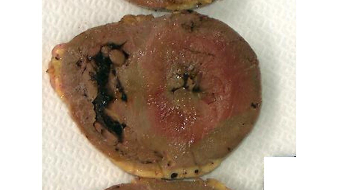
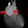
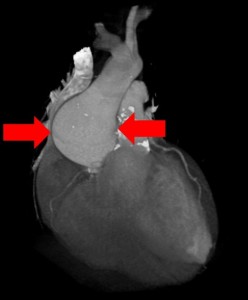
The top image shows the heart sac opened in the chest. The sac is full of blood (cardiac tamponade). The bottom image shows the heart after the blood around it has been removed. The view is of the back of the heart. There is a T-shaped hemorrhage. The right coronary artery (RCA) wraps around from the front and is seen on the top right. The left circumflex coronary artery (LCX) also wraps around from the front and is seen the top left. The posterior descending coronary artery (PDA) branches off the RCA and travels down the middle of the back of the heart.

What was the story here?
The patient was a middle aged man who came in to the hospital short of breath for several days after getting through a cold. He had a coronary artery stent put in his right coronary artery. The procedure seemed to go well. Later in the afternoon, he became dizzy, collapsed and died. His wife spoke with the cardiologist who explained it was probably a “stroke.” The wife could not understand why, suddenly, there was a stroke right after the procedure. She also didn’t believe he had any blockages because the husband had been athletic. She requested an autopsy and retained a lawyer.

What caused the blood on the back of the heart?
The blood on the surface of the back of the heart tracks along the coronary arteries. (The coronary arteries travel over the surface of the heart). This suggests the blood came from the coronary artery itself. Because a stent was placed in the same area as the hemorrhage, this suggests that a coronary artery ruptured related to the placement of the stent. Other possibilities would be a CPR-related contusion (but that would not track along blood vessels); or a tear of the muscle heart muscle wall (but that did not happen either).

How did the person die?
After the vessel ruptured, the blood also leaked into the sac around the heart. When the heart sac fills with blood, there is no room for the heart to beat. The heart is squeezed by the blood in the sac. This can cause the patient to collapse and die. This is what happened here.

Could the rupture have happened on its own, unrelated to the procedure?
No. Spontaneous rupture of coronary arteries is documented but extremely rare. The rupture was right in the region where the stent was placed and indicated the procedure had a role in the rupture.

What else did the autopsy show?
The autopsy showed very little coronary blockage overall and a viral infection of the heart muscle (viral myocarditis).

How did the autopsy help?
The autopsy helped on a variety of levels. It determined the cause of death (coronary rupture with tamponade). It also provided information on the prior health of the heart (viral myocarditis). The patient’s shortness of breath was likely from his heart infection; and his heart infection was likely a complication from his recent viral illness (“cold”). The results also shed light on the role of the procedure in the patient’s death. Because the patient’s coronary arteries were “open,” the procedure was unnecessary. Moreover, it caused his death.

What was the impact of the autopsy results on the family’s emotions?
Difficult as the information was to consider that the death was intimately related to the care, the information from the autopsy helped the family move on from the painful restlessness of “not knowing.” At the same time the results deepened the wife’s concern regarding the cardiologist’s explanation and behavior. She began to suspect that her husband had been left to die at the time of the code (because the cardiologist did not seem to consider a diagnosis related to the heart). On top of this, she felt profoundly dismissed that there seemed to be no consideration that her husband’s death may have had any connection to a procedure hours before — a “common sense” consideration given the time frame. The autopsy results validated her concerns. Before the autopsy, the wife was left not knowing what had happened with the cardiologist telling her it was a stroke. After the autopsy, the wife knew what happened, which diminished her dependence on the cardiologist and seemed to have a liberating effect.

Why would the cardiologist suggest the cause of death was a stroke?
The cardiologist’s comments are filtered through the wife and we can’t really know what he said. If the report is accurate, it is concerning. Was this “physician hubris” (“My procedure can’t have caused a problem. I know I did everything right!”)? Was he being dismissiveness of the wife’s intelligence? Was this fear of a medical error being discovered and an attempt to deflect the wife from the “truth”? Was he really suspicious of a stroke on clinical grounds? Which option, or some other, cannot be ascertained here. But, the possibility that truth dismissed caused the patient’s death (by inattention to a procedural complication during the code) is horrific.

Summary Comment:
The issues in this case are multiple. They include a missed initial diagnosis (myocarditis); an erroneous assessment of coronary blockages (open and not blocked); a procedural complication (coronary artery rupture related to stent placement); and, possibly, misdiagnosis of stroke during the patient’s code. Probably least in question here is the actual technique of placement of the stent — the reason being that the coronary artery wall into which the stent was placed was not firm with plaque, but a pliable normal vessel. It was possibly more easily damaged by a metal stent than one shored up by at least some firm cholesterol plaque. While seemingly remote, there is a possibility that standard of care was met at various points. It would take a review of the presenting data, angiograms, the operative note, the post-operative care, and so on. However, the autopsy served to secure the truth — of benefit to the clinicians and to the wife sorely in need of information.

Sleuth It – The Case Without Findings

[hupso url=”http://autopsychicago.com/sleuthitdecember2015/”]
Click here to skip to discussion.

Test tube photo

A 44 year old man dies suddenly after a night of “partying.” The coroner has made her evaluation but declined to perform an autopsy. The two sons request an independent autopsy, which you perform. The body is thin, but well nourished. There is no evidence of trauma and the autopsy exam is entirely normal. You save some blood for toxicology, even though the family has not requested testing.

Afterwards, you meet with the sons. They explain that their father was drinking beer and doing “shots” of hard liquor, but wasn’t really “that drunk.” You talk though the pluses and minuses of testing the blood for alcohol: the sons already know their father was drinking, so the test wouldn’t add that; but the test might give a clearer sense of how much alcohol was “in the system.” The sons don’t want any toxicology testing, saying they know “how hard” their father partied.

Suddenly, they turn to one another, whisper, and then turn back. “Oh, yeah, our father also ….”

Which of these disclosures would help in this case?

Their father:

a. Had high blood pressure
b. Was depressed and took the antidepressant Zoloft
c. Did cocaine
d. Had a serious family history of high cholesterol
e. Just started taking Coumadin (blood thinner) for a problem with his heart rhythm.

Stay tuned next month for a discussion of the case.


January 9, 2016 Update:
Case Discussion.

It would help to know that the father used cocaine (choice c.). Cocaine can cause spasm (clamping down) of normal coronary arteries, block blood flow in the artery and cause a heart attack that way. It could certainly have caused the death.

It may also help to know if the father had an abnormal rhythm (choice e.). That might be explored more to see in what way the abnormal rhythm might have been a risk factor for sudden death.

Coumadin is a risk factor for bleeding into the head — but you performed the case and there was no bleeding. So the part of option e. that is helpful is the mention of an abnormal heart rhythm, not the use of Coumadin.

High blood pressure is a risk factor for a large heart and an abnormal rhythm — but you performed the case and the heart was normal.

Family history for high cholesterol is a risk factor for high cholesterol and blockages — but you performed the case and there were no blockages.

Zoloft is not commonly a drug that causes overdose.

[hupso url=”http://autopsychicago.com/sleuthitdecember2015/”]

Monthly Case: Aortic Rupture

June Monthly Case - safe  view

Background: The case is of an elderly man with sudden onset of left hip pain. He was diagnosed and treated for arthritis, but died two weeks later. The body is oriented diagonally in the photo. The head would be at the upper left and the feet at the lower right. The star shows the inside of the rib cage. The heart and lungs have been removed. Most of the abdominal organs have already been removed. What happened? What caused the man’s symptoms? Why did he die?


Slide2

Discussion: The patient’s aorta ruptured just above where it branches near the pelvis. This branching is called the aortic bifurcation. The aorta ruptured (black oval) towards the left hip, sending blood (curved arrow) towards that side of the body and causing the patient’s discomfort around the hip.

Isn’t aortic rupture serious? Why didn’t this patient die right away from bleeding? The patient did not die suddenly because the rupture was partly contained inside the nearby tissue. The blood did not freely enter the abdominal cavity — which could have been an immediately life threatening event. Instead, the blood tracked and pushed into the pelvic tissue, the upper leg tissue and around the hip. The patient was mistakenly thought to have had arthritic pain, but the pain was, in fact, from the pressure of blood in the tissue around the hip.

What caused the rupture? The surface of the patient’s aorta (outlined in red) is rough and not smooth. This tells us, the patient’s aorta was covered in cholesterol build-up (plaque), a risk factor for the rupture. The aorta was also widened in this area (difficult to see in this view), so the patient had an aneurysm which ruptured.

Below is a diagram of the anatomy we are talking about.

Slide3

Why did the patient die? The patient did not lose enough blood to die from bleeding. Instead, he developed an abdominal infection on top of the rupture and that caused him to die.

Sounds of Death – Airway Mucus Plugging

Diagnosis: Airway mucus plugging

[one_half] Photo Case 1 Feb 2014
[/one_half] [one_half_last]


[/one_half_last]

What is shown?
The photograph shows the large airway opened from behind. Seen are: the voice box (left, black arrow), trachea (center); two mainstem bronchi (right, white arrows). The airway is completely filled with mucus.

How did this happen?
This patient was very weak (from cancer and from an infection), had advanced Alzheimer’s disease, and could not “clear” the mucus. Normally, people can “clear” mucus by coughing it out of the airway (and then swallowing or spitting it out). The patient was too weak to do this. So the mucus built up and blocked the airway.

How did this patient die?
The mucus blocked the airway and gradually choked the patient, preventing airflow into the lungs.

How did the autopsy help the family?
The autopsy findings allowed the family to address specific, emotionally-charged and distressing medical-related issues:

The patient’s level of comfort. The family noticed gurgling sounds during breathing and was concerned this meant the loved one was uncomfortable or suffered near the time of death. The autopsy confirmed the presence of mucus but cannot specifically comment on suffering. However, an understanding of the specific effect of mucus on air flow helped the family feel more comfortable that the patient may not have suffered.

Here’s how that worked. Because mucus builds up gradually, the patient must have had a gradual decrease in air flow (rather than abrupt, as in sudden choking). This suggests the likelihood of a gradual and long term decrease in body oxygen. This means the brain likely also had a gradual decrease in oxygen supply. Low oxygen levels cause the brain to lose or decrease consciousness. An unconscious person cannot experience suffering. This means there was a high likelihood the patient did not suffer.

The patient’s level of care. The family worried that, with all that mucus, the patient should have been suctioned by nursing staff. The autopsy cannot comment on treatment options. However, by discussing the issues and findings, the family can consider alternate points of view. For this patient, the following questions had to be considered: Given her terminal condition, what were the treatment goals? Was full care the treatment plan? Or was hospice in place? What palliative measures (e.g. suctioning) were agreed upon? Was the treatment plan made clear to the family or, more importantly, made by the family?

A family that agrees to hospice but requests suctioning suggests an active and human struggle in accepting imminent death and letting go of the loved one. A family that requests full care (including suctioning) and does not see it suggests a different set of issues.

Summary.
In this particular case, the autopsy allowed the family to come to terms with the terminal nature of the cancer; understand that their observation (of breathing) reflected the process of dying; focus their energy on bereavement rather than anger (over perceived nursing issues); and thereby achieve a sense of peace.

[button label=”Latest News, Events and Articles” url=”/” target=”_blank” color=”#f05134″]

Sleuth It: Case of Tendon Repair (Part 1)

Tendon writst

A family calls to request an autopsy. They complain that a recent surgery “killed their mother.” The patient had a history of heart disease, high blood pressure and diabetes and lived alone. The surgery was a tendon repair of the wrist and took place three days before the patient died. The surgery reportedly went fine and recovery was going well — just with some pain at the surgical site, but no swelling or infection. The family visited regularly. On the third day in the evening, the family was with their mother when she passed out.

What questions do you have for the family now?
At this point, what do you think could have made the mother pass out?

Submit your comments here and stay tuned for Part 2.

Monthly Case: Early Graft Closure

Safe with notes
The case was of a middle aged man who died suddenly two weeks after a surgery. One of the photographs shows the cause of death. The other photograph from the same patient shows a similar process, but was not the cause of death.

Case discussion:
The photographs show grafts to the heart. These were grafts from a coronary artery bypass procedure. The grafts came from a vein in the leg (saphenous vein) and were attached to the heart to allow blood flow around blocked coronary arteries. The grafts have been cut open to see along the length of their insides.

The bottom graft shows the cause of the death. This graft is completely blocked by the clot seen at the right. The clot appears as a red tube inside the graft. It blocked blood flow from getting completely through the graft to where it needed to go – the heart muscle. It was as though the heart was functioning without this graft at all.

This was a sick heart with a recent serious heart attack that prompted the bypass surgery emergently. The graft was critical to the patient’s health and life.  Without the open graft and the blood flow to the heart, the patient died.

The top photographs shows a small amount of clot healing onto the inside of the graft.  It’s not enough clot to block the blood flow in the graft, but is the same process. This clot is brown and not red because it is starting to heal and scar, which changes the color.

To the left of the clot in the top photo is a hole surrounded by small sutures.  This is called a “side anastomosis.”  This graft was used twice.  Blood flow passed through the side anastomosis to a blood vessel there; and blood flow passed through the end of the graft (all the way at the right) to a blood vessel there.  Often, grafts can be used to bypass more than one blocked vessel.

Early graft closure from clot (thrombosis) is a known, but rare complication of coronary artery bypass grafts.  It’s unrelated to the technique of the surgery or any intraoperative factors, but just a part of the body’s reaction that can sometimes happen to the graft itself.

Meaning to the family:
In this case, while it was helpful to know that the graft closure was “no fault” of the surgeon, the patient had tell-tale signs of trouble during recovery after hospitalization (shortness of breath and fainting) that were not attended to. This was a complicated post-operative story to sort though. The autopsy was helpful to the family by clarifying the cause of death and its relationship to natural processes, the surgery and post-operative care.

Monthly Case – Uremic Pericarditis

Feb case safe view The case was of a middle aged man treated with intravenous antiobiotics for a skin and muscle abscess which developed after an operation. The patient died suddenly months after the abscess healed. The normal organ is seen on the right.

Discussion: The patient developed kidney failure as a result of toxicity from intravenous antibiotics. The patient required short term dialysis. He was recovering from his kidney failure when he died suddenly. The metabolic and possibly immunologic consequences of kidney failure had taken a toll on the heart, although the kidneys themselves were recovering. Uremic pericarditis is a known but poorly understood complication of kidney failure. “Uremic” means “related to kidney failure.” The heart is covered in fibrin (a blood protein) and inflammation and had begun to scar onto the surrounding sac (pericardium). “Pericarditis” means “inflammation of the pericardium.”