Category Archives: Blog

Monthly Case – rupture AV fistula – redo

AV Fistula safe view

What is shown?
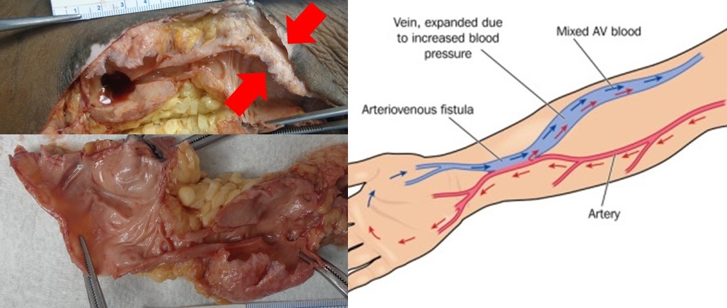
The upper left photograph shows a dissection of an arm. The AV fistula is visible below the skin. The upper arrow shows how paper thin the skin is right next to the AV fistula. The lower arrow shows how paper thin the AV fistula itself is.

The bottom left photographs shows the AV fistula removed from the arm. Each attached clamp holds an expanded portion of the AV fistula. The connection in between is less expanded. The yellow tissue is attached fat.

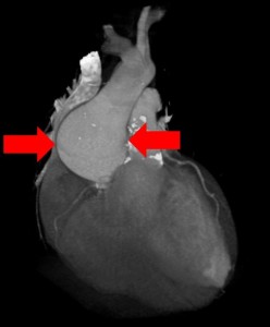
The right image shows a diagram of an AV fistula.

What is an AV fistula?
The “A” in AV fistula stands for “arterio” (relating to arteries – the high pressure blood vessels carrying blood away from the heart). The “V” in AV fistula stands for “venous” (relating to veins – the low pressure blood vessels returning blood to the heart). A fistula, in this case, means a surgically created connection. So an AV fistula is a surgically created connection between an artery and a vein. It stands for arteriovenous fistula.

Why did this patient have an AV fistula?
AV fistulas are created by surgeons to help patients who need dialysis. To have dialysis, large needles need to access the blood stream. An AV fistula provides a large vein to allow for this kind of access.

How do AV fistulas provide large veins?
Veins are normally used to the low pressure blood flow inside them. If a vein is surgically connected directly to an artery (under higher pressure), the vein swells and expands under the pressure from the artery. The vein becomes big enough to be used for dialyis. This swollen vein is illustrated in the image on the right.

What was the story here?
In this case, an elderly man needed dialysis for his kidney disease. He had an AV fistula placed. The fistula worked for many years. One month before he died, his fistula began to leak. The first time was at home. He woke up to find his bed sheets soaked in blood. He went to the hospital, received blood transfusions and was sent home. A couple weeks later, he bled again, also at home. A third time occurred during dialysis, requiring his arm to be tournequetted while the bleeding was stopped. He was sent home again. The following week, he was found in the bathroom having bled to death from his arm.

Why did the family request an autopsy?
They wanted to know more information about why the AV fistula leaked. In addition, the family wondered why their loved one, after several bleeding episodes, did not receive any specific plan to prevent further bleeding (for example creating and using a new dialysis access site). It seemed that the only medical care was to replace lost blood, but that the AV fistula continued to be used. They were looking for more information to understand what had happened.

What did the autopsy show?
The autopsy showed why the AV fistula had become at risk for rupture and bleeding. The photographs show how thin the skin was over the AV fistula, how thin the AV fistula was itself and how close the two were. After multiple punctures, the two had scarred together, thinned and become brittle. This explained how the fistula was at risk for rupture and bleeding.

How did the autopsy help?
Despite the clear clinical history of bleeding from the arm, it was not clear to the family what had happened. They imagined that the fistula itself was full of many, many holes (from “too many” needle sticks). Instead, the autopsy showed that most of the AV fistula was normally formed; and that the problem was right at the surface where the skin had thinned and become brittle over the years. This provided a clearer, less “dramatic” (and, therefore, less stressful) picture for the family.

In addition, the discussion allowed for a validation of the family’s concern about continued use of an AV fistula that had ruptured three times. For an AV fistula that had worked for many years to rupture more than once, there would be no reason to think the bleeding wouldn’t happen again. Unfortunately in this case, the patient died as a result. Talking it through with a clear understanding of the findings helped the family come to terms with events surrounding the loss of their loved one.

[one_sixth]
[/one_sixth][one_sixth]
[/one_sixth][one_sixth]
[/one_sixth][one_sixth]
[/one_sixth][one_sixth]
[/one_sixth][one_sixth_last]
[/one_sixth_last]
[one_third]
[/one_third][one_third]
[/one_third][one_third_last]
[/one_third_last][one_third]
[/one_third][one_third]
[/one_third][one_third_last]
[/one_third_last]
Autopsy Center of Chicago provides compassionate, comprehensive caring service to families in need. Talk to you us about your concerns. We are here for you.
We will guide you through the process, talk you through your concerns and work to bring you to a place of peace.

Learn more about our services here.

Cancer’s hidden stronghold: your identity

You have cancer. You loved one has cancer. You are in a cancer support group. You give to cancer research. You want a cure for cancer. You join a cancer bereavement group. Cancer impacts your identity.

Cancer sure has a lot of power over your life. Cancer, the diagnosis, may be inside you or your loved one’s body. That’s Mother Nature’s deadly mischief. She does have that power. But cancer, the identity, is a choice. How are you participating in that choice? How free does society make it for you to realize it’s a choice?

Even before prognosis and treatment options rise to view, the toll of cancer’s uninvited diagnosis resounds through the senses. Society is ready for you, even if medicine isn’t. There are groups and associations and runs and friends with stories and family reactions and clinical trials. Come on in — we’ll make your fight or acceptance or palliation as comfortable as possible.

You become part of a community with resources. But is the subtext that your experience and identity also belong inside this community? Patients and families with cancer can understand patients and families with cancer.

There are benefits to having a community. Cancer associations and support groups serve as good will centers to support cancer research. They allow for a communal rallying cry to put a stop to the terrible disease. That’s an important goal. They also foster trust and a feeling of membership. This balances the “wrecking ball” of cancer that destroys lives and, often, relationships. A cancer community is also a way to share resources and allow for learning about about the biology and treatment of disease — there’s an important learning curve each patient must progress through to navigate their health care choices.

But there’s a cost. When you enter the cancer community, the door may lock behind you. Who says that your experiences can be understood only by those who have shared in the diagnosis? That gives too much credit to cancer.

Cancer, the identity, comes out of modern medicine. Imagine a time before cancer could be diagnosed. How would we manage our experiences — witnessing an enlarging neck, extreme weight loss, or a change in thinking? Knowing the diagnosis helps. But the experience itself takes processing. There’s a broader community available for support and understanding. Engaging — presuming access to and acceptance from — a broader community diminishes the power of the diagnosis and destroys stigma.

Here’s a present-day example. There’s a group of patients with lung cancer who die in a most shocking and dramatic way. The tumor can sometimes rupture a large blood vessel in the chest. Imagine blood everywhere — on the floor, on the wall, on the loved one. It can look like a crime scene, prompting police inquires and confusion in the family. Especially if there’s no knowledge or teaching (you, provider!) that this type of death might happen and why, witnessing such an extreme death can be traumatic. It can, therefore, also be isolating.

Who can understand this extreme experience? Others who lost a loved one to cancer? Or the sister of an alcoholic who watched as her brother’s esophageal varices rupture to make a similar scene? Or the son who saw his father with diverticulosis and rectal bleeding? Take away the diagnosis and they’ve all seen the same thing.

What of the young spouse who lost her partner to gastric cancer shortly after marriage? The unique journey of a millennial through the health care system — Did the doctors know how to tell her it was terminal? Was there anyone her age in the support group? — might resonate more with another millenial sharing a journey of terminal illness than with an elderly woman who lost her husband to gastric cancer after a lifetime.

There are children isolated from participating in family illness (and then grief) because cancer was the “family secret.” No one would acknowledge that “daddy had colon cancer.” Does their adult support best come from other patients with colon cancer? Or from another individual who’s family would not explain “daddy had heart disease”?

Why, cancer, do you get to keep me from sharing the humanity of my trauma by virtue of this medical diagnostic word?

You don’t. Not with us.

We, at Autopsy Center, are creating “I was there” groups. It’s to cut cancer down to size and stop cancer from defining human identity. The medical diagnosis causing the experience is being confused with the experience itself. That’s giving cancer way to much power.

Everyone who goes through the cancer journey with their loved one has a unique experience with unique themes (delayed diagnosis, abdominal pain, multiple family losses to the disease, and so on). Similar experiences can be found in patients with heart disease, stomach ulcer and Alzheimer’s disease.

It’s the double edged sword of modern medicine. The diagnosis lays the path for treatment. The label functions to organize community funding for research. But there needs to be self-awareness that all these facets are in play.

Let science define the cells. Let research and medical resources fund diagnosis-specific groups. But let society be inclusive. Let humanity transcend the diagnosis. We can find support in many places.

This is the goal of Autopsy Center’s “I was there” groups. These are on-line communities for people sharing common end-of-life experiences. None of them are defined by a specific diagnosis, say … cancer. You can guess some of the group titles: “Massive Hemorrhage,” “Terminal illness was the family secret,” and “My partner and I were both young.”

It’s to know there’s someone out there who saw what you saw in that room, was treated the way you were treated in the hospital, felt what you felt seeing your family cope. It’s to share the humanity of the experience. Cancer can’t tell you how to do that.

Live Stream Autopsy in the Time of Fake News

There’s nothing more real than an image of an autopsy. You’d think. Not in the time of fake news.

We started live streaming autopsies in 2017 to hundreds of thousands of viewers (our first drew in over a million). Family consent beforehand and maintaining patient confidentiality during the case are, of course, requirements. We have an open Q & A during the case where I talk with viewers and answer questions. The videos are left up after the case and more Q & A rolls in, for later discussion. It’s been a phenomenon for us.

To sort it through, I pay attention to where our viewers lead us. Sure, we get our share of horror fans and, shall I say, worse? Our videos, I’m told, have found their way into a “serial killer” site. I’m not sure what that means. I’m hoping it’s for people CSI-curious about criminals…not a club for serial killers. And, through Google’s truth-outing, I’ve recently seen that we’re now up on a site called Cute Dead Guys — it is what it sounds like. I only know because of brief descriptors out there. I have not (and will not) be seeking the password access that tens of thousands have. To get personal, many of you who know me will have discovered the shocking truth that I don’t like horror movies or watch CSI. When I’m not doing autopsies, it’s Toy Story and Moana (“See the light where the sky meets the sea, It calls me, And no one knows how far it goes…”). Ahh, Disney. But I believe passionately in compassionate education about death, dying, health and the body. So I do what I do. It’s a balance.

On a more tame scale, our viewers have a fascination with death. Our social media serves an important normalizing function there. Our comment section on Facebook is filled with statements like “I watch all your videos. I love them. My boyfriend thinks I’m a weirdo.” And then a stranger replies: “I love them, too.” And so friends are made and it becomes ok to be interested and to learn. We work for that comfort on our site. It’s a place for people to feel comfortable with themselves and the topic. I explore and understand the body after death to help the family with closure. The autopsy is an intimate medical procedure. We design the Facebook page to highlight respect for the process and for our families. But we see the struggle with taboo in our culture that makes it so hard for people to be ok with wanting to know.

Next seems to be the largest group of our viewers: one that simply has a huge need for knowledge. Seeing the inside of the body is educational. Many comments reflect this: “I didn’t know fat was yellow.” “The skull is so thick.” “That’s where the pituitary is — no wonder they talk about some tumors being ‘inoperable’.” Many people aren’t exposed or educated about what’s on the inside. We show that. It fills a need.

And then, we have our share of professionals — nurses, paramedics, funeral directors and a variety of students. “Better than a textbook” is a theme. Stay tuned for “augmented reality.”

So that’s an encapsulation of who views us. It’s a simplification — we get moms who watch with their 3-year-old; one who watches to relax before going to sleep, and others. We think carefully about what happens to the material once we put it out there and consider how we should make it available. But we believe in education and keep our “graphic warning” front and center for our videos. The idea is not to shock, but to educate for those interested.

Now onto our place in the time of fake news. I wonder if one of our draws is simply that we are real — if that brings in a good number of our 23K Facebook followers. We are real but not a “reality show.” With the near-impossibility finding the truth on-line these days, I’m wondering if we become a place for that. In addition, we foster trust by talking without taboo. Our discussions cover death, dying, the body, nudity, health care, social media issues and the truth itself. Our Facebook page is modeled after the meaning of autopsy, “to see for one’s self.”

Let me add another phenomenon from our page. We have many people asking if the person in the video is dead. How can that be? We are removing a brain. This person in the video cannot be alive. The answer is complicated. In part, it’s the shock and surprise of seeing the procedure on-line. We’re not used to seeing autopsies. We’re used to surgeries — health care for the living — so it’s an adjustment. Another reason has to do with a deeper psychological readiness to understand “dead.” Like children developmentally, not all adults are there. It can take some processing. I get why people ask. And lastly, as one insightful Facebook fan commented — thinking of the person as alive is just easier. It can be psychologically stressful to be faced with full-on “dead.”

Only rarely do we have anyone call out our work fake. I have seen it only twice in closely reviewing thousands of comments. One person commented on Facebook that the fat looked fake — this was easily explained because there is no bleeding in an autopsy (since the heart stops circulating blood). So the fat sits there as a bright yellow plasticky-looking layer. The second comment, from YouTube was more direct: “Clearly fake. Pasta. Props on the special effects”[sic].

The draw to our page, value to our viewers for the education, the struggle some have with “understanding dead” and occasional call-outs of Fake! point to one thing: a person’s psychological readiness for the truth determines what they look for and can accept. I suspect this is true in the media in general. We should have compassion for people who need fake news. They must have some deep limitation as to what they can handle from the daily, painful reality that bombards and overwhelms us in the media. (This, of course, does not mean we should tolerate efforts to undermine the truth.) On the flip side, those fighting to access the truth — or place to say a five-minute autopsy video is of interest — show courage. Fake news is a broad cultural expression of psychological stress in the face of reality.

Autopsy Center is here to develop a place for those ready (or who can be ready) for the truth. One of the things we are doing at the Center is developing our high school program, Live from the Morgue. Schools from across the country can join us simultaneously during one of our video-educational sessions. We’re developing our school-to-school shared experiences specifically with an eye to breaking barriers at an early age.

Join us when you’re ready and if you’re interested. We’re here to share the truth. To our pasta-commenter from YouTube. No, not fake, not pasta, not special effects. Real. The human body. Props on how beautiful life has made us.

Check out our videos on Facebook or YouTube or download our Autopsy app.

Reflection: Live from the Morgue

Small group of university students working on computer in a library. Young people finding information for their college assignment.

Live from the Morgue
is our live-feed interactive video educational experience for high schools.  
 
To learn more or register your class, please click here or contact us.


The voice on the the screen flickered silent as I leaned in.

“I can’t see you. I can’t hear you,” I said into my still computer, hoping — maybe at least I was coming through on the other side.

“Call me back,” I offered, not knowing if the connection in Georgia was as useless as mine in Chicago.

Today I was going to use my scalpel in front of thirty high schoolers. Each had signed up to watch the autopsy. I show everything – from Y-incision to bowel splay. It’s for education, I had explained. We had had a chance to talk about it beforehand. The older students were protective of the younger ones. It’s not for 9th graders, they said.

“Too immature. Too young,” they explained. How is it that sophomore high school students feel so strongly about students just one year younger?

But perhaps the were right. There were no 9th graders at today’s event.

“Why not?” I had asked just before losing signal, still looking to understand.

“Too wimpy,” one student offered. The freshmen had opted out on their own.

A bubbling beep focused me back on the screen and the teacher’s familiar smile met me.

“Hi! I see you now,” came the drawl from across the country. “We’re ready for you!” he said. Our connection was back on.

I saw the last few students shuffling in behind him, head and legs cut off by the limits of the webcam lens. As they sat, their faces floated down into view over their pale desks, eyes on me. Each, as instructed, sat with juice bottle and cracker snacks — our plan to ensure students don’t pass out during the experience.

“Ready?” I asked, poised for the first cut.

The video count down 5-4-3-2-1 flashed across my computer and streamed across theirs. My blade entered the patient’s left shoulder. It swiftly passed down to the chest and the yellow fat below filled the split. There was, of course, little blood because the patient was dead.

I explained everything. The bruising on the ribs was from CPR. The pockets in the colon were called diverticulosis (not infected). We were waiting for toxicology testing — I had shown them an active case. I always protected the confidentiality of the patient, but our library of family-authorized video footage was growing. We updated our video sessions frequently.

After the event, we processed. Peering from a thousand miles away into this classroom, the students at first seemed indistinct. Slowly they came to life — the student who wanted to be a nurse; the student who was “grossed out” by the colon; the student who recounted her own experience with a dead body — one she did not know. Others took steps to solve the case. “When will toxicology testing come back?” many asked. The curiosity and connection were palpable.

Next year’s session can’t come soon enough, I thought as I logged out with Georgia.

Maybe we’ll even see some freshmen.

Essay: The Only Acceptable Option

IV image

Joe and his wife, Carla, talked about it when she got the news – she didn’t want chemo. She didn’t want chemicals to destroy her body. She made that clear to Joe. Her father went through something awful when he got cancer and she just didn’t believe in those poisons. And they had done their research — how much does chemotherapy actually work for cancer of the stomach?

There was only one acceptable option – treatment had to be “natural.” They considered a trip to Mexico for some “alternative” treatments but money was a concern. So they sought out doctors nearby who believed the same as they did — that cancer should be treated without western poisons. After a few weeks, they found their medical sanctuary and the treatments began.

Carla seemed to be doing well on weekly intravenous hydrogen peroxide, Joe explained as we sat in my office. I swallowed and could feel the blood rushing from my face. What kind of doctor treats cancer patients with injections of hydrogen peroxide? My heart sank. This is why I help families — vulnerable in so many way and at such critical times.

Joe’s faith in the “treatment” and his disbelief in the outcome were profound. It was stomach cancer but Carla had been eating — maybe a little trouble swallowing but not much in the way of other symptoms. The tumor must not have been growing, he had concluded. The treatment was working. His older brother, Mark, was with us, nodding and supporting him, listening and asking questions.

What went wrong? Joe wanted an autopsy to find out.

It showed widely metastatic cancer. Outsized lymph nodes formed a slowly tightening noose around the esophagus; a destructive bio-lava of tumor pushed from the stomach into the pancreas; and metastatic growths nested insidiously throughout the liver. Remarkably, the gastric tumor had grown out and away from the stomach, not into the stomach itself. No wonder there wasn’t much in the way of swallowing or digestive symptoms. But this apparent health belied the tumor’s silent and dangerous growth nearby.

When we met again to discuss the results of the autopsy, Joe was shocked. She had seemed to be doing so well. He was surprised that her tumor was so advanced. With diagrams and details, I went through the case step by step.

He was stunned, but understood. And then he understood what this meant — that the treatment had not worked. And then he understood the inevitable — that he had participated in the choice of treatment.

“Did we do the right thing? Should we have gone with chemotherapy?” he asked, wrestling with guilt as so many families do.

I sorted through the question, looking for a way in to help. It wasn’t my judgment to pass. Gastric cancers do not respond well to chemotherapy. Choices at the end of life are important and personal. But he had said “we” and Carla had really made the choice herself with Joe supporting her — it really hadn’t been his decision. She had been adamant. Maybe I could point that out?

And while Carla seemed to have driven the choice of “treatment,” it wasn’t clear to me how deep Joe’s mistrust of chemotherapy was. Many chemotherapies do work. Hodgkin’s lymphoma, for example, can be cured. I thought it could be worth exploring. What if Joe had such a choice to make again? What if that choice was for himself? Feeling protective of him, I thought I could explore it gently and carefully. Autopsies are about the truth and can be turning points for families.

But his brother spoke first, injecting the only acceptable option he could see — that there could be no second guessing.

“Of course you did. Of course you did,” he said, shooting a glance at me and touching his brother’s arm.

It was an older brother’s protection. The dynamic here was powerful.

“Yes,” Joe nodded, losing himself into his brother’s verbal embrace.

I thought Joe could have handled more, and wondered where the conversation might have gone. I grimaced internally as I saw the opportunity dissolve. It was now too risky to start a discussion that could be misconstrued as “blame.” “Do no harm” — my physician oath ingrained — meant that I could not risk infusing guilt on top of grief.

So I said nothing. It was the only acceptable option.

New Procedure: Small Incision Autopsy

Small Incision Autopsy

We’re now offering an autopsy whose sole purpose is to provide families with information about inherited conditions: The Small Incision Autopsy.

Our over-riding goal is to provide families with closure after a loss. Generally, this means understanding why the patient died. But it can also mean explaining what may have caused troubling symptoms, assessing changes related to procedures and other treatments, and teaching about basic disease processes. If the death is unexpected and sudden, families can also wonder if there is an inherited condition to worry about. An autopsy can sometimes help by finding out.

The Small Incision Autopsy uses a small incision — much smaller than that of a routine autopsy — to access important areas of the body that can harbor just the information that could help the family.

Right now, we’re focusing on heart disease — a main cause of sudden death. Some important inherited causes of sudden cardiac death can include coronary blockages from high cholesterol; and the very under-reported valve condition, bicuspid aortic valve. Taking a focused look at the heart can uncover these and other heart conditions. The family can then get tested and treated. Knowing can save lives.

[button label=”Learn more” url=”/small-incision-autopsy” target=”_blank” color=”#f05134″]

Sleuth-It Diagnosis: Bicuspid aortic valve

[button label=”Read the Sleuth-It” url=”/sleuth-it-what-counts-in-the-heart” target=”_blank” color=”#f05134″]

Safe bicuspid

What counts in the heart? The number of parts to each valve counts. In this case, the number of parts (leaflets) of the aortic valve was important.

You stopped counting after seeing two aortic valve leaflets. Normally there are three.

Two aortic valves rather than three is an inherited condition called “bicuspid aortic valve.” It affects 1-2% of the population.

Bicuspid aortic valve can be associated with aortic aneurysm and rupture, typically in late middle age among males with the condition.

The inheritance is not well-understood but does not have to affect every family member.

Once this diagnosis is suspected, family members can get tested. If any family member has the condition, they can be monitored for disease progression and treated (with blood pressure medicine, surgery if needed, and so on).

Knowing the diagnosis can save the lives of the affected family members.

The autopsy helped with the family’s closure; but also did much more than that by alerting the family to an inherited genetic condition.

Here’s more information about bicuspid aortic valve.

Sleuth It — What Counts in the Heart?

Aortic aneurysm imageYou perform an autopsy on an elderly man who died suddenly at home. The gentleman was stoic, somewhat distant from his family and never sought medical care. There are four adult children who call you to perform an autopsy. They would like to know what happened.

During the autopsy, you find that the sac around the heart is full of blood. Your first thought is to consider either a heart attack with rupture of the heart muscle; or an aortic aneurysm with rupture of the aorta.

The coronary arteries are whistle clean (no significant blockage by plaque) and the heart muscle is healthy. There was no heart attack. But the aorta coming out of the heart is twice as wide as normal and there’s a half-inch tear in it close to the heart. It’s the aorta that has ruptured.

You consider high blood pressure as a common cause of aortic aneurysm. But if there were many years of high blood pressure, the heart should be bigger than normal and the kidneys should have a rough surface. The heart is normal size and the kidneys are smooth. It doesn’t look like there was high blood pressure.

You wonder about the inherited condition, Marfan syndrome, which goes along with a stretched out aorta and aortic rupture. But patients with Marfan syndrome tend to be tall and this gentleman is short. Marfan syndrome is unlikely.

You continue a systematic assessment of the heart, continuing with the heart valves. First you assess the valves on the right side of the heart: Tricuspid valve. “One, two, three leaflets. All thin and pliable. No problem there,” you think. Now the pulmonic valve. “One, two, three leaflets. All thin and pliable. No problem there.” You move on to the left side of the heart, looking at the mitral valve. “One, two leaflets, no problems.” Then you look the aortic valve, “One, two….”

Suddenly you realize you better call the family in. The autopsy results may save their lives.

What did you see?

[button label=”Click here to see if you solved the case” url=”/sleuth-it-discussion-1″ target=”_blank” color=”#f05134″]