Category Archives: Blog

Monthly case – Congestive Hepatomegaly

Congestive hepatomegaly Oct 2014 Safe

Diagnosis: Congestive Hepatomegaly

What is shown?  The photo shows a markedly enlarged liver (seen on the left).  The liver is about twice as big as normal.  Also shown is a normal liver (seen on the right and roughly the size of a football). You can see from the rulers that the scale is same in both pictures.  The colors are different because the liver on the right was embalmed before the autopsy while the liver on the left was not embalmed.

How did the liver get so big?  The liver got big because it filled with blood.  The extra blood was entirely responsible for the large size of the liver.  The blood was inside the liver’s blood vessels.  The liver cells themselves did not get bigger nor did they increase in number.

How did so much blood get into the liver?  The blood got into the because this patient had heart trouble.  When the heart stops functioning well (can’t pump blood forward), the blood can back up into nearby organs, for example, the liver, lungs and spleen.  This causes these organs to swell.  That’s what happened in this case.

What does “congestive hepatomegaly” mean?  Congestive (or congestion) is the term for “blood vessels filled and swollen with blood.”  “Hepato” means liver; and “megaly” means “big.”  So this is a “big liver from blood vessels swollen and filled with blood.”

What does this finding mean when seen during an autopsy?  This finding often is part of the process of dying or it can mean the heart had been sick for a while.  It’s important to know a bit more about the story to understand how to make sense of a congested liver.  While very noticeable during an autopsy, the finding does not really give much information about the cause of the heart problem, or even when that problem started.  It just indicates either that the heart had a problem or that the heart failed as part of the process of dying.

What was the story here?  This was an elderly woman who had a complication after a minor surgery on a limb.  She was in the hospital for many weeks after the surgery and then died.

What was the family’s question here?  The family was angry.  They wanted to understand how, if at all, the treatment or surgery may have caused their loved one to become sick and die.

Comment long hospitalizations.  Long hospitalizations are particularly challenging to sort through during an autopsy.  This is because, over the course of the hospitalization, many things happen to the patient.  This makes it hard to figure out – just looking at the body – what problems were there before the patient was hospitalized; what problems developed possibly as a complication of a treatment; what changes indicated healing; and so on.  These cases require close study of the chart, x-ray studies, and the history.  And the answers aren’t always clear.

How did the autopsy help?  In this case, the family was desperate to make sense of what happened.  Understanding which findings (like the liver enlargement) where the result of the process of dying and which caused the death were important to sort through.  Doing that allowed the family to come to terms with the medical issues and their loved one’s experience after her surgery.

Monthly Case: Second lung cancer

Safe Lung cancer

What is shown?  The picture shows a large tumor (oval) of the left lung.  The tumor is about 3 inches wide, from top to bottom. The tumor is right next to the main airway (arrows).

How did this patient die?  The lung cancer grew into the airway and blocked this patient’s breathing.

What type of tumor was this?  Tumors in the lung (and any organ) come in different types. This one was a “small cell carcinoma.”  It’s called this because of the small size of the cells seen under the microscope.  This is a very aggressive (fast growing) lung tumor.  Prognosis is commonly six to twelve months, even with treatment.

What was the clinical story here?  This patient was an elderly woman who had been a smoker and had emphysema.  She had had lung cancer in the other lung – the right lung – about ten years before. That right lung tumor was completely taken out by surgery and was not thought to have spread (metastasized) anywhere back then.

Why did the family request an autopsy?  The family wanted to know why there was a tumor in the left lung if the surgery “got it all out” from the right lung ten year ago.  This was confusing to them.  They began to feel mistrustful of how first treatment went.  Did they really get it all out?  Were they being dealt with honestly?  How could there be a second tumor in the lungs now?

What did the autopsy show?  The autopsy showed the past surgery on the right lung. About a third of that lung had been removed. There were normal healing changes and no “left over” tumor on that side.  The left lung tumor was found (described above).  There were two metastases in the liver and metastases in the bone marrow.

How did the autopsy help?  The autopsy allowed for a discussion of the biology of tumors and gave a perspective on the patient’s care.

Here were the possibilities to consider:

-That the second tumor was part of the first because some was, in fact, left behind after surgery ten years ago and had just kept growing.
This could not be.  Because small cell carcinoma is so fast-growing, if there were any tumor left from the surgery, it would have grown quickly at that time.  It would not grow so slowly to take ten years to come to medical attention.  Also, the tumor was in a completely separate lung.  Any tumor remaining on the right side would have grown in the right lung not the left.

-That there were tiny metastases at the time of the first surgery that were undetected but then grew and showed up ten years later. 
This is also could not be, again because small cell carcinoma grows so quickly it would not take ten years for metastases to come to medical attention.

That the new tumor was a complete different tumor from the first.
This can only be what happened here, because the above possibilities were ruled out.  Also, it is known that once a patient gets one lung cancer, they are at increased risk of a second, different lung cancer. It is something that is known to happen.

How did the discussion impact the family?  The information staved off anger towards the original surgeon.  The family could rest at ease knowing that the original surgery had, indeed, accomplished its medical goals.  The family did not need to feel any concern for any past medical mismanagement, error or dishonesty because there had been none.  Past treatments were completely unrelated to the new tumor.  This was an entirely new and different tumor.  The family had not known this could be a possibility.

The discussion also provided a reminder of the risks of smoking. 

Lastly, learning more about the biology of lung cancer gave the family a language and context to understand and talk about their loved one, each important in the grief process.

HB5348 – Crib Bumper Pad Ban

Senate Floor Pano

By Rabiya Bilfaqi, 2nd year law student, DePaul University College of Law
and Ben Margolis, M.D.

HB5348 Ban Crib Bumper Pads
Name of Sponsor: Representative Emily McAsey (D)85th District
Date Sponsored: 2/10/2014
Status: Pursuant to Senate Rule 3-9(b) / Referred to Assignments
Synopsis As Introduced
Amends the Children’s Product Safety Act. Prohibits a commercial dealer, manufacturer, importer, distributor, wholesaler, or retailer from selling, offering to sell, leasing, or offering to lease a crib bumper pad in the State. Imposes a civil penalty of not less than $100 and not more than $500 for each violation. Provides for the deposit of these civil penalties into the Attorney General Court Ordered and Voluntary Compliance Payment Projects Fund.

Introduction
Accidents are among the leading causes of infant deaths nationally.  In Illinois in 2010, 55 out of 1116 infant deaths were from accidents.1  The importance of cribs and play environments is highlighted by product recall statistics.  In the last two years, the US Consumer Product Safety Commission has recalled more than 5 million cribs, bassinets and play yards.  Illinois House Bill 5348 seeks to eliminate accidental infant deaths due to one such product – crib bumpers pads. The bill amends the Child Product Safety Act and bans the sale of this product entirely.

What is the Child Product Safety Act?
The Child Product Safety Act regulates products that pose safety hazards to children. The Act covers car safety seats, children’s toys, and children’s furniture. The crib bumper ban pertains to the last category.

What is a crib bumper?
A crib bumper is a set of four pillow-like pads that are tied to the inside of a crib above the mattress.

What do we know about crib bumper dangers?
A baby can suffocate when wedged against a padded crib bumper or strangle by a bumper tie around the neck.  Between 2008 and 2011, the National Center for Child Death Review received 14 reports of infant suffocation in which a bumper was relevant in the death.2 A study in the Journal of Pediatrics (2007) looking at coroner and medical examiner reports of 27 accidental infant and toddler deaths concluded that “[crib bumper] use prevents only minor injuries. Because bumpers can cause death, …they should not be used.”3  The American Academy of Pediatrics notes that “there is no evidence that crib bumper pads protect against injury, but they do carry a potential risk of suffocation, strangulation or entrapment because infants lack the motor skills or strength to turn their heads should they roll into something that obstructs their breathing.”4

History of Crib Bumper Pad Legislation
In 2010, the Chicago Tribune reported that federal regulators failed to warn parents that crib bumper pads pose a suffocation risk to infants even though they knew about the hazard.5 Subsequently, the Food and Drug Administration reported the known risks associated with bumper pads including suffocation. In 2011, Chicago became the first city in the United States to ban the sale of baby crib bumper pads. This helped set the stage for the statewide ban on crib bumper pads and helped to raise awareness of the suffocation risk that crib bumper pads pose. There is an ongoing effort nationally to implement crib safety laws. In 2013, Maryland became the first state to enforce a complete ban on the sale of crib bumper pads. States like Texas, California and New York all have general crib safety standards, although they do not yet have laws specific to crib bumpers.

What does a safe crib look like?
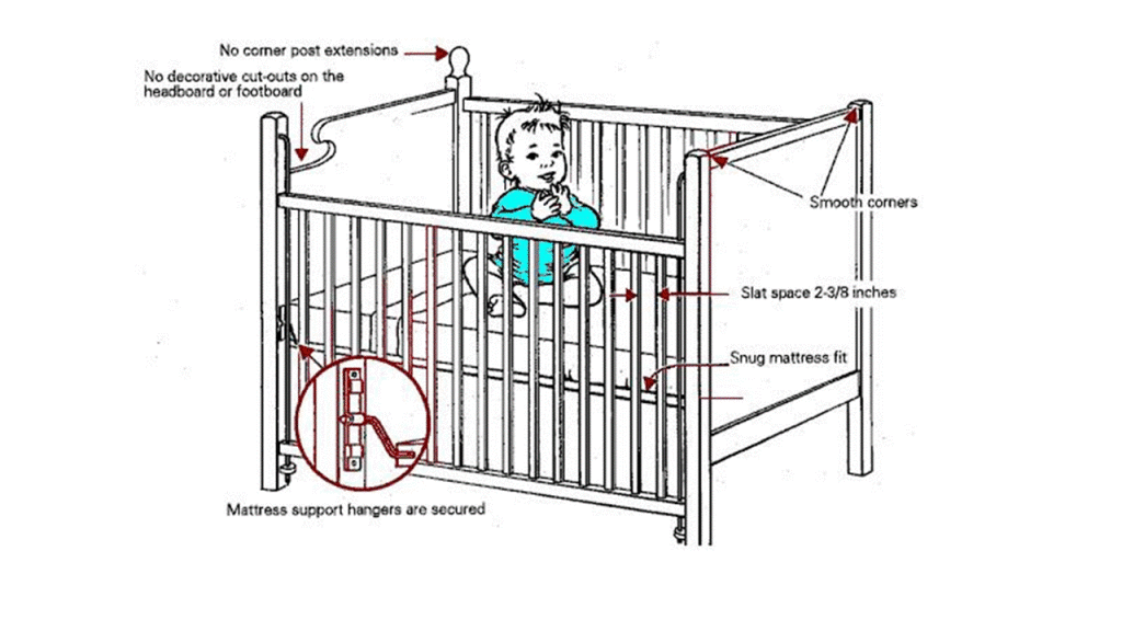
Many states classify a safe crib as one that meets the following safety standards6 :
•smooth corner posts that extend 1/16-inch or less above end panels
•slats narrower than 2-3/8 inches apart
•a secure mattress support that does not release easily from corner posts
•no cutout designs on the end panels
•no tears in mesh or fabric
•no cracked or peeling paint to prevent lead poisoning
•no missing or loose screws, bolts, or hardware
•wood surfaces that are smooth and free from splinters, splits or cracks; no sharp edges, points, or rough surfaces

Crib Safety

Comment on Sudden Unexpected Infant Death Initiative
Medical examiners and coroners have moved away from classifying deaths as Sudden Infant Death Syndrome (SIDS). They are more likely to classify deaths as accidental, suffocation-related or with an unknown cause. SIDS is now being called Sudden Unexpected Infant Death Syndrome (SUID). The Center for Disease Control (CDC) states that “inconsistent practices in cause-of-death determination hamper the ability to monitor national trends, ascertain risk factors, and design and evaluate programs to prevent these deaths.”7 The CDC’s research on SUID focuses on efforts to improve data collected at infant death scenes and to promote consistent reporting of cause and manner of death for SUID cases.

Final Comment
At Autopsy Center, we know what families go through when they experience a loss.  And, in the world of grief, parental loss is profound.  Our goals is not only to support families during a loss, but to work to prevent such losses.  We support public health initiatives, such as HB5348, which save lives.

Resources
Product Safety Recalls
Resources for Bereaved Families

References
1 Leading Causes of Infant Death 2010. Illinois Department of Public Health website http://www.idph.state.il.us
2 http://www.childdeathreview.org/home.htm
3,4 Thach, Bradley T., George W. Rutherford, and Kathleen Harris. Deaths and Injuries Attributed to Infant Crib Bumper Pads. The Journal of Pediatrics 151.3 (2007): 271-74.e3. Web.
5 Gabler, Ellen. Federal Regulators to Study Safety, Suffocation Hazard of Crib Bumpers. Chicago Tribune., 12 Dec. 2010
6 http://www.freecasereview.com/InjuryLawArticles/cribsafetytips.htm
7 CDC’s Sudden Unexpected Infant Death Initiative. Centers
for Disease Control and Prevention
. 10 May 2011. Web. 7 Sept. 2014.

Monthly case: Fungal abscess

Fungal abscess protected

What is shown?   The photograph shows the right and left lungs and the unopened sac around the heart (pericardium, dashed line) still inside the chest.  The arrows point to a region of pus in the right lung.  It’s a bit smaller than the size of a playing card and not quite half an inch deep.  This is an abscess.  The white flecks are fungus (mold).  This is a fungal abscess.

What is the clinical history here?  This case is of an elderly man with numerous bouts of “pneumonia” and unexplained fevers over several months. Each time, the patient was treated with antibiotics and improved, but the “pneumonia” always came back.  The “pneumonia” was first diagnosed by CT scan.

How did the autopsy help the family?  The autopsy provided an explanation for a situation that had been a mystery.  The family was distressed and confused as to why treatment did not work.  The autopsy provided the “big picture” here – that, underneath it all, was an abscess, not a pneumonia.  This provided relief to the family, and a sense of peace on one level.  Now they knew why their loved one never got better.

Why didn’t the abscess heal with antibiotics?  Antibiotics reach areas of infection by travelling through the blood stream.  An abscess is a pocket of pus with no blood flow inside it – antibiotics can’t easily get inside.  The treatment of an abscess is to drain it surgically.  A lung abscess that does not get drained will sit in the body and continue to cause symptoms (fever, breathing problems, general weakness, etc.) or, as in this case, death.  In addition, the patient received antibiotics (which treat bacterial infections).  The patient needed antifungals because this was a fungal infection.

How did the family manage with this new information?  While the family was surprised and relieved, they were angry that the abscess had not been detected prior to the patient’s death.  Specifically, they wondered why it was not detected on the initial CT scan.  Also, they were angry because they had asserted themselves with providers, questioning the diagnosis and care, but were dismissed.

How did the family discussion proceed from this point?  In this case, it was very important to make sure that feelings of guilt were not added to the family’s anger; and to help them manage their anger by providing information and perspective.  Specifically, it was not the family’s fault the diagnosis was not made.  It was important to value the family’s efforts during the time they cared for their loved one, validate their commitment to their loved one, and emphasize the complexity of the health care system.  Also, it was important to give the family as many tools as possible to interpret the situation.  For example, regarding their concerns about the first CT scan, the possibilities were that:

-the abscess was not there at that point (e.g., that it developed later)

-the abscess was there but difficult to see for some reason related to its appearance on the CT (e.g., perhaps related to the abscess being relatively flat)

-that the abscess was there but was was missed by the radiologist

While the last possibility was the family’s concern, a discussion of the biology of abscesses and the limitations of radiology studies helped sort through the issues.  The discussion provided tools to the family in thinking about the experience, and helped them formulate questions when seeking information from the providers.  Fungal abscesses are notoriously difficult to diagnose and often missed.

Summary.  By providing concrete information from the autopsy examination and by engaging the family on an educational level, this allowed the family to feel a bit less helpless and a bit more empowered moving forward during their vulnerable and difficult time.

What’s Wrong with You – Fiction

What's wrong with you image

What’s Wrong with You

by Ben Margolis

Ars Medica Vol 10, No. 1 (2014), p. 142-146

Author’s note: caution, graphic content


“What’s wrong wit’ you?” asked the shopkeeper.

“Ain’t nothin’ wrong with me,” scowled Mr. Jackson as a flap of skin unfolded across his chest. Hinged at the side like an armoire door to Mr. Jackson’s body, the flap opened at the breast bone, curling outward towards his left arm….

Read the full piece:

What’s Wrong with You (pdf)

What’s Wrong with You (on-line)


Monthly Case: Blocked stent

July safe

What is shown? The upper left photo shows a metal mesh tubing. This is a stent. It’s about half an inch wide and a few inches long. This stent is inserted into a blood vessel to help keep the vessel open and the blood flowing (illustrated on the top right). The bottom photo shows the vessel in a patient who had an autopsy. In this case, the type of vessel is a vein. It is a large vein that drains the blood from the leg.  The vein, is cut into smaller pieces to see what’s on the inside. You can see metal stent as a circle of dots that that presses up on the inside of the vessel. The inside of the stent is filled with yellow solid tissue. This is scar tissue. The vessel should have been open and and empty.

Did the scar tissue cause a problem?  Yes.  The vessel was completed blocked by the scar tissue, so it could not drain the blood from the leg.  The left leg was swollen.  There was blood return from the leg, but it was through smaller side veins, and not enough to keep the leg from swelling.

Did the blockage cause the patient to die?  No.  The patient did not die from this.  He lived with a swollen leg.  The swelling developed gradually over many years (see below).

Why was the sent placed? The stent was placed in the vein to keep it open. A blood clot had formed in the vein, blocking flow. The stent at first helped with that by opening up the vessel to allow blood flow back from the leg.

Why did the scarring happen? The scarring happened because, even after the stent was put in, more clot formed inside the vessel.  The body’s response to a clot is to heal the clot by forming scar tissue. It is a natural response of the body to any clot that forms inside a vessel.  The process of forming this scar took many years.

Does the scar means there was a problem with the procedure or with the stent? No, stents are not perfect. Any time “foreign” material (like metal) is put into the body, there is a known risk that the body will have a reaction to it. Also, this patient was already at risk for forming clots (see below). There is always a “risk-benefit” decision for any procedure. The thinking is like this: Without the procedure, the patient would have had blocked circulation in the legs. With the procedure there is the risk down the line that the stent could get blocked. The best decision was to do the procedure to help with the “immediate risk” to the patient from his own blockage the first time he had a clot.

What was the story here? This was an 18 year old man with a disease related to sickle cell anemia called with S-C disease. “S” stands for “Sickle”. “C” is a genetic variation of sickle cell anemia. These patients are typically a bit less sick than patients with sickle cell anemia but also have serious problems.

How did this patient die?  This patient died from “Acute Chest Syndrome.”  This is a condition that happens commonly in S-C disease.  Here’s how it works:  In S-C disease (just as in Sickle Cell Anemia), the red blood cells can clump and block the blood flow in many areas of the body.  When that happens specifically in the bone marrow, parts of the bone marrow can die.  The dead bone marrow leaks into the blood stream, travels up to the lungs, and the little clumps of bone marrow block the circulation there.  This causes the patient to die.  Unfortunately, many clinicians are not aware of this complication of S-C disease.  This patient’s clinician did not consider this diagnosis.

How did the autopsy help?  The autopsy helped the family come to terms with the loss.  The family also chose to share the results with the treating physician which served to educate him about this condition.  It was an empowering move by the family in a situation that was otherwise marked by helplessness.  The specific finding of a blocked stent was incidental to the cause of death but facilitated a general discussion of the patient’s medical past.

Reflections on an Autopsy Seminar

[one_half]

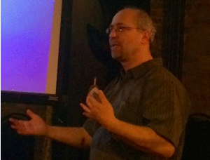
Life Matters Media photo credit.

[/one_half] “Was that a one-glass slide? Or a two-glass slide?” I called over to an attendee, gently teasing, as I advanced to the next photo in my presentation. He was all but hiding in the corner. This was the one who had brought in a bottle of wine to our cabaret-style theater where we were holding the event, Science to Humanity: The Autopsy. He had come accompanied by some friends, but really a glass of wine (or two, or three, or a bottle) is what he thought he would need to get through experience. I was just checking in on him.

He smiled.

“I’m good!” he called out.

I was glad to know our at-first-fearful gentleman was doing well. Our attendees always do.

People really don’t know what to expect when they come to an autopsy class. For most, that’s just right. What they get is so different from what they ever imagine.

The class is respectful, paced gradually, engaging and set with a warm, open tone. Who would have thought that the room often fills with laughter? It’s the laughter comedic improvisers know comes from the place, “I recognize that from life.”

This makes sense in a class about the autopsy, perhaps not so paradoxically. The autopsy is about closure. And closure is human. This is a class where people connect.

I have been giving this class since 2012 and we are delighted to now be moving to the International Museum of Surgical Science. It is a milestone for us. We are honored to be able to hold the event in such a respected venue, and looking forward to continuing our outreach.

The class began on the 84th floor of the then Sears Tower — my office space at the time. Our first attendees were a young couple, and that was it. The young man had called me in seeming desperation hours before.

“I missed the ticket deadline! Can we come?” he inquired.

“Of course,” I responded, and called the office to reschedule the room reservation I had just cancelled.

And so, with a bit of a hurry, the first class was held with the three of us. Slide after slide, question after question, proceeded; explanations turned into stories; and, as it always does, the class became an experience. The man ultimately disclosed that his mother had had the same procedure I had shown in one of my slides — placement of a gastrostomy tube. The slide showed a medical mishap. I gasped, fearing what he might infer about his mother’s experience, and I quickly reminded him that I see cases only when things “go wrong” — it’s just the nature of my work. When I was done reassuring him — emphasizing the countless times the procedure had helped patients — he sighed and relaxed. And the flow of questions continued. The young couple walked out hand in hand.

While the class is educational and not a counseling experience, I am aware that what happens there can have an impact. I see this in the letters I receive from attendees and in the reviews.

One attendee wrote months after coming to the class:

“I just wanted you to know that your workshop has been helping in our understanding that [our mother] might not have felt any pain [during her death].”

And a recent reviewer commented:

“I wasn’t considering myself “in bereavement” when attending [the class] although I did lose someone very close to me earlier this year….I was incredibly suprised at how some of the presentation struck a chord with me and answered some questions that I didn’t even realize that I had. I left feeling a sense of peace that I didn’t realize I needed. I didn’t know that certain questions could even be answered.”

So I take the class seriously, keep it “light” — as much as possible for this topic — but respectful and am open to the human experience as well as sharing my own. People leave satisfied.

Our gentleman with the bottle of wine told me at the end that the slides were his favorite part.

“They really weren’t bad,” he said. “I wanted to see some more.”

Next event: July 30. Tickets/information/reviews: here

Poem: Invitation

Med Humanities (British Medical Journal) 2013;39:72 doi:10.1136/medhum-2012-010312

Invitation
Ben Margolis

Come.

I will show you grief.

Look here.

You want to see?

The open refrigerator door frames the lovers.

Just this way.

Follow me.

We’re almost there.

You will stand with me by the side.

You will have to look down—it’s private.
[one_half] But can see from the corner of your eye.

A cold visitation deep in the quiet maze of the hospital basement. Just myself witness. Best man to half an alter-vow fading under caresses. Now you along side.

‘Can I cut a piece of hair?’ she asks, no longer knowing what was hers. Crush hold memento.

‘Of course, or we can take care of that for you’. I would start the case as soon as she finished. An intrusion of strong gloved hands and calculating blades waiting.

The hair was her final quiet request, but the visit had started with a scream. Mrs. Paznyk had seen her husband immediately after he had died, but not since, now a day later.

‘Close his eyes! Close his eyes! Close his mouth! Stop it!’ Eyes as wide as his, mouth not quite, body pulled back. She was unprepared for rigor mortis’s play. Swift sculptor of all dead. No embalmer’s rewinding art yet.

‘It’s part of death’.

‘Ok. It’s part of death. It’s part of death. It’s better this way. It’s normal’. Recoiled pain and suffering stuffed into calmed adult readiness. But still bruising, tearing under the surface.

We had agreed on 15 minutes for this last viewing. It made sense to have a start and an end, or she would have remained there, in the cold walk-in, drinking passionate memories and tender commitments from a bottle of Amontillado-laced grief.

I shall leave you here.

If this is what you were looking for.

Unless you would like to meet four year old Javier.

Rolling onto my lap.

‘Read it again!’

Giggle.

‘Ok. I love David. He’s such a bad boy!’

Booga-booga-booga belly!

Nose up to mine.

Smiling.

Hand on book.

‘Read it again!’

You start.


Footnotes

Competing interests None.  Provenance and peer review

Not commissioned; internally peer reviewed.

Author note:  All names and events are fictional.

[/one_half]

New event venue

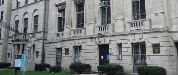
Our event, Science to Humanity:  The Autopsy, is moving to a new venue. Join us at the International Museum of Surgical Science starting July 30 for Science to Humanity; and stay tuned for more educational events in 2014-2015. The facility is located along Lake Shore Drive in the Gold Coast just one block south of North Avenue. Discounted parking is available nearby, including at the Chicago History Museum. The venue is wheelchair accessible.

IMSS PHOTO

While at the museum you can see some of the most innovative tools and procedures from the history of medicine. Exhibits cover such diverse subjects such as ophthalmology and orthopedics, and come from all over the world and every part of history. The IMSS collection ranges from an Iron Lung and early artificial limbs to a 19th century pharmacy featuring Dr. Miles’ Cactus Compound, Lydia Pinkham’s Blood Medicine and many other elixirs from ages past.

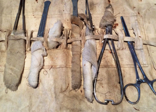
Surgical tools

In addition to artifacts from medical history, guests can also see modern artwork devoted to medical innovation and the creative spirit of health science.  Works by Artist-in-Residence Vesna Jovanovic and the Anatomy in the Gallery exhibit provide a current perspective on the long tradition of art in medicine.

Vesna Jovanovic

The IMSS was established in 1954 by the International College of Surgeons and Dr. Max Thorek. It is the oldest medical museum in Chicago and one of the few in the Midwest. Capably staffed by volunteers, the museum has become an attraction for students and adults alike.

International Museum of Surgical Science
1524 N. Lake Shore Dr.
Chicago, IL 60610
312-642-6502

Monthly case: Heart Rupture

 
Covered Heart Rupture June cropped 2014

What is shown?   The top photo shows the sac around the heart (pericardial sac) cut open and lifted to expose the sac filled with blood (star).  The right lung is seen on the right.  On the bottom left is the heart.  A clamp has been inserted into the site of rupture.  The tip of the clamp (actually inside the heart) is illustrated with dashed lines.  On the bottom right is the clot that had escaped from inside the heart into the pericardial sac.

What was the history here?  This was a functional, active elderly woman who was days earlier released from the hospital after a heart attack.  She collapsed at home.  Paramedics were called and performed CPR, but she could not be resuscitated and died.

Why did the family request an autopsy?  The family was concerned that the mother died shortly after hospital discharge.  They wondered if the hospital had released her “too soon” (i.e., that she should have stayed in the hospital for her safety and monitoring).

What happened here?  The heart broke open (ruptured) causing blood to escape into and fill the pericardial sac.

How did this happen?  This rupture was likely caused by CPR in combination with a weakened heart muscle wall from the patient’s heart attack.  Cardiac rupture from CPR is a rare but known complication of this life-saving procedure.  Rupture of the heart wall is also a known complication after a recent heart attack.  The two can at times go together.

What else can cause the heart muscle to rupture?  Sometimes the heart muscle thins as it scars after a past heart attack from months or years prior.  With large heart attacks, part of the heart muscle can scar to “paper thin” in some patients.  This thin wall can rupture even without CPR.

Why, in this case was CPR considered as a cause of the rupture? In this case there was considerable trauma to the chest wall and other areas of the heart that suggested the CPR was vigorous.  Specifically, there were broken ribs on the left; and blood in the soft tissue underneath the breast bone.  Lastly, there was bruising on the back of the heart.  Since CPR is performed from the front, the force must have been strong enough to mash the back of the heart on back of the chest and cause this bruising.

How did the autopsy help?  The finding of heart rupture from the autopsy was new information for the family.  Families often wish to know if “everything” was done to save the patient.  Since heart rupture is severe and often causes death, it was clear that resuscitation efforts would not have resulted in “bringing back” the patient.  “If only” concerns (e.g., “if only they had worked on her a little longer”) could be laid to rest as continued CPR would not repair a rupture.  The autopsy also served to exclude other unanticipated causes of death (e.g. pulmonary embolism) and validated the family’s concerns regarding the heart itself.  Thus, the autopsy’s definition of the medical issues allowed the family to move forward. Lastly, considering the complex processes here (the patient’s disease, hospital care, hospital discharge planning, cause of the collapse, timing and need for CPR, and heart rupture) allowed for a thorough discussion of the medical issues – always important in the bereavement process.

Note:  CPR is and remains an important and life-saving measure.  CPR-related heart rupture is rare.